

Assessment first
Every mole is examined before removal. If any feature suggests it needs analysis, that's explained to you and the removed tissue is sent for laboratory examination. Most results are entirely reassuring, and having the answer is preferable to wondering.

How it's done
The method depends on the mole's type, size, depth, and location. Shave removal suits raised moles sitting above the skin surface and generally leaves a small flat mark. Surgical excision is used for deeper moles or where the full thickness needs removing for analysis, and involves stitches. Your dermatologist explains which applies and why before proceeding.

What to expect
The area is numbed, so the procedure itself is comfortable. It's usually quick. Aftercare involves keeping the site clean and protected, with specific instructions given on the day.

Scarring
Any removal leaves some mark. The realistic outcome is a small scar that fades over months and is less noticeable than the original mole, not no mark at all. Where a mole is in a cosmetically sensitive area, we discuss the likely result before you decide.

Regrowth
A shave-removed mole can occasionally regrow. Surgical excision is more definitive.

Why people have them removed
They catch on jewellery and collars, get irritated by shaving or clothing, become sore if twisted, and are cosmetically bothersome when visible. Removal is usually straightforward.
Why they appear
Friction is the main factor, which is why they cluster in areas where skin folds. They become more common with age, and are associated with weight and, in some cases, with insulin resistance, so a sudden crop of new tags is worth mentioning at consultation.
How they're removed
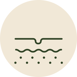
Small tags are removed quickly in clinic. The method depends on size and location; snip excision, cautery or cryotherapy are all used. Multiple tags can generally be treated in the same appointment.
What to expect
Local anaesthetic is used where appropriate. Healing is quick, usually with a small scab that resolves over days.

Recurrence
Removing a tag doesn't stop new ones forming elsewhere, since the underlying tendency remains. New tags in the same area are common over time.

Types we treat
Common warts, usually on the hands and fingers. Plantar warts (verrucae) on the soles, which grow inward under pressure and can be painful to walk on. Flat warts, which appear in clusters, often on the face or legs. Filiform warts, which are thread-like and often appear around the face.

Why they persist
Warts can resolve on their own, but this often takes a long time, and meanwhile they spread. Plantar warts in particular tend to be stubborn, since pressure drives them inward.
How we treat them
Cryotherapy or freezing is the most common in-clinic treatment and usually needs several sessions spaced a few weeks apart. Cautery or minor excision may be used for resistant warts. Prescription topical treatments can be used alongside, particularly for larger clusters.

Realistic expectations
Warts frequently need more than one treatment session, and can recur because treatment removes the wart but the virus may remain locally. Several sessions is the norm, not a sign that something has gone wrong.

Preventing spread
Avoid picking or scratching, which spreads the virus to adjacent skin. Cover plantar warts in shared facilities. Don't share towels or footwear.
Why a biopsy might be recommended
To identify a lesion whose nature isn't clear from examination. To confirm a diagnosis where a rash or condition hasn't responded to treatment. To distinguish between conditions that look alike but need different management. Or to establish the nature of tissue removed during another procedure.
How it's done
The area is numbed with local anaesthetic. Depending on what's needed, the dermatologist takes a shave sample from the surface, a punch sample using a small circular instrument, or an excisional sample removing the lesion entirely. It takes only a few minutes.
Afterwards
Some biopsies need a stitch or two; others heal with a simple dressing. Aftercare instructions are given on the day.
Results
Samples go to a laboratory for analysis. Your dermatologist explains the findings and what they mean for your treatment.
Why it's worth doing
A biopsy converts uncertainty into an answer. Where treatment hasn't been working, it frequently explains why and changes the plan to something that will.

Cysts
Epidermoid and sebaceous cysts are benign lumps beneath the skin, most often on the face, neck, back and scalp. They're usually painless, but can become inflamed, infected or tender, and often grow slowly over time.
Why complete removal matters
A cyst is a sac. Draining it relieves it temporarily, but if the sac wall remains, the cyst refills, which is why cysts that have been squeezed or drained repeatedly keep returning. Definitive treatment removes the sac intact.

Timing
An actively inflamed or infected cyst is usually settled first before excision, since operating on inflamed tissue gives a poorer result. If a cyst is currently inflamed, treatment may happen in two stages.
Other benign lesions
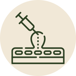
Lipomas, soft, benign fatty lumps beneath the skin, and other small benign growths can also be removed in clinic, depending on size and location. Larger lesions may need referral, and we'll tell you clearly if that applies.
How it's done
Under local anaesthetic, through a small incision, with the lesion removed as completely as possible. Stitches are usually needed and removed at a follow-up visit. Removed tissue is sent for analysis where indicated.

Scarring
Excision leaves a scar proportionate to the incision. Your dermatologist will discuss the likely result relative to the lesion's current appearance before you decide.